


- Meet Carol*
- Clinical Assessment†
- Imaging Report
*Hypothetical patient.
The objective of Amyvid image interpretation is to provide an estimate of the brain beta-amyloid neuritic plaque density, not to make a clinical diagnosis. Image interpretation is performed independently of a patient’s clinical features and relies upon the recognition of unique image features.
†Image interpretation should be performed independently of the patient’s clinical information. The use of clinical information in the interpretation of Amyvid images has not been evaluated and may lead to errors.
‡Amyvid PET scans are interpreted independently of the patient's clinical information.
The objective of Amyvid image interpretation is to provide an estimate of the brain beta-amyloid neuritic plaque density, not to make a clinical diagnosis. Image interpretation is performed independently of a patient’s clinical features and relies upon the recognition of unique image features.
 70 YEAR-OLD
ART TEACHER―Widowed Since 2012
70 YEAR-OLD
ART TEACHER―Widowed Since 2012 ENJOYS PAINTING and being with her 3
grandchildren
ENJOYS PAINTING and being with her 3
grandchildren Arrives
30 mins late for her visit, seems anxious
Arrives
30 mins late for her visit, seems anxious Describes
her morning as "CHAOS"
trying to get her grandchildren to school
Describes
her morning as "CHAOS"
trying to get her grandchildren to school- Reports
more difficulty with memory for the past 2 years and asks, "THIS IS JUST NORMAL FOR MY AGE,
RIGHT? "
- Repeated the same story about her
morning twice
- MMSE
26/30

 Complete history and physical are
normal,
Complete history and physical are
normal,
including neurologic exam—reveals no focal deficits Routine lab tests normal, as well as
TSH, B12, and folate
Routine lab tests normal, as well as
TSH, B12, and folate Structural
MRI is read as "age
appropriate atrophy"
Structural
MRI is read as "age
appropriate atrophy" Diagnosis is persistent MCI: Follow-up
scheduled in 2 weeks—family member is requested to attend
Diagnosis is persistent MCI: Follow-up
scheduled in 2 weeks—family member is requested to attend An Amyvid PET scan is ordered to differentiate diagnosis and decide, along with Carol, on appropriate management

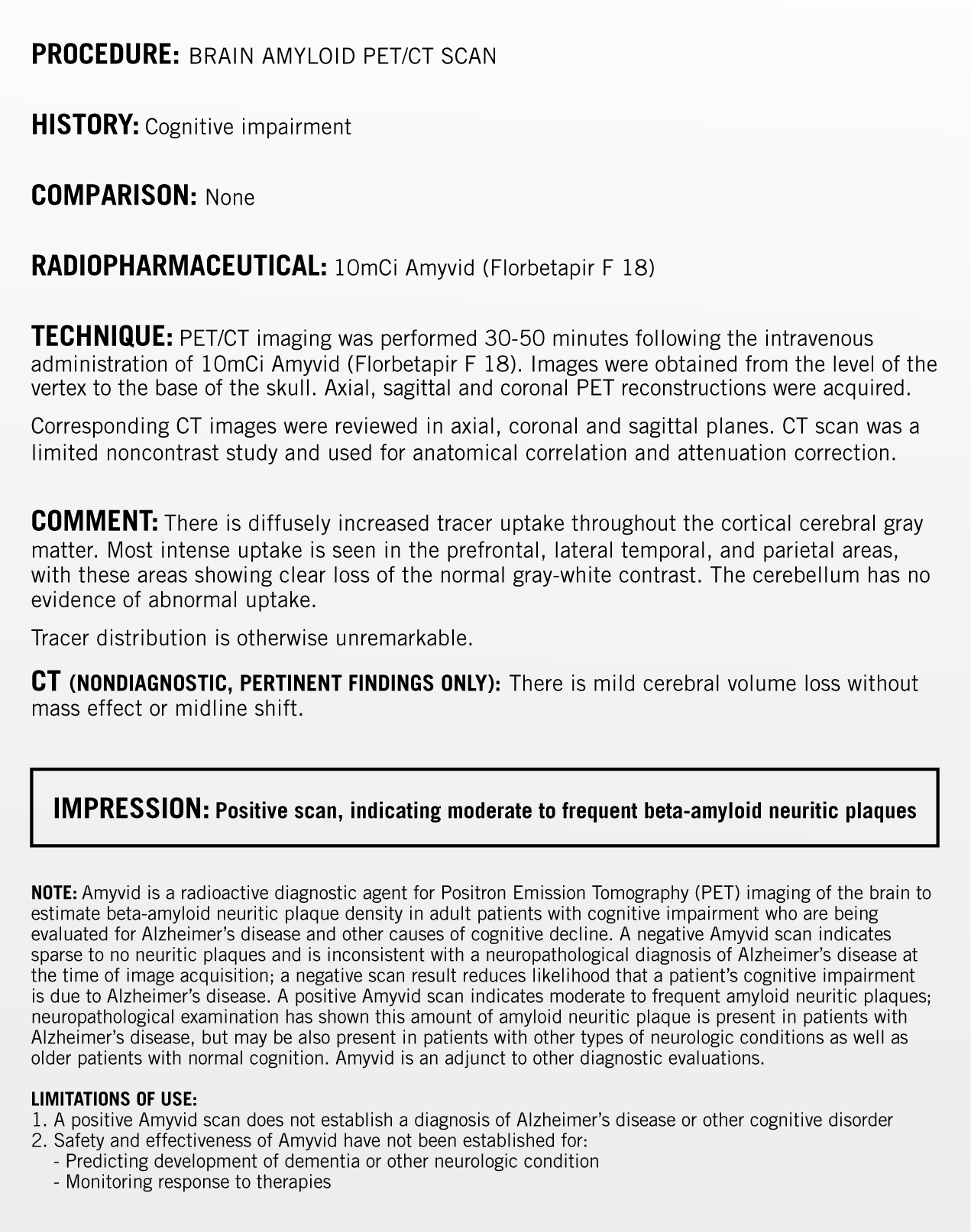
Example of a positive Amyvid PET scan imaging report.
POSITIVE scan‡
- Indicates presence of significant (moderate to frequent) amyloid neuritic plaques1
- Consistent with a neuropathological diagnosis of AD1
- Neuropathological examination has shown this amount of amyloid neuritic plaque is present in patients with AD, but may also be present in patients with other types of neurologic conditions as well as older people with normal cognition1
- Amyvid is an adjunct to other diagnostic evaluations. A positive Amyvid scan does not establish a diagnosis of AD or other cognitive disorder. The safety and effectiveness of Amyvid have not been established for predicting the development of dementia or other neurologic condition, or for monitoring responses to therapies1


- Meet George*
- Clinical Assessment†
- Imaging Report
*Hypothetical patient.
The objective of Amyvid image interpretation is to provide an estimate of the brain beta-amyloid neuritic plaque density, not to make a clinical diagnosis. Image interpretation is performed independently of a patient’s clinical features and relies upon the recognition of unique image features.
†Image interpretation should be performed independently of the patient’s clinical information. The use of clinical information in the interpretation of Amyvid images has not been evaluated and may lead to errors.
‡Amyvid PET scans are interpreted independently of the patient's clinical information.
The objective of Amyvid image interpretation is to provide an estimate of the brain beta-amyloid neuritic plaque density, not to make a clinical diagnosis. Image interpretation is performed independently of a patient’s clinical features and relies upon the recognition of unique image features.
 71 YEAR-OLD
ACCOUNTANT with cognitive concerns
71 YEAR-OLD
ACCOUNTANT with cognitive concerns ACCOMPANIED TO DOCTOR’S OFFICE
by his wife of 40 years- Wife
describes his
PROGRESSIVE CHANGE OVER THE PAST 3 YEARS- Gradually lost ability to work with numbers; his wife now manages their finances
- Used to enjoy lively dinner conversation; now he is less talkative
- His memory is worse, but sometimes recalls nearly everything
 GEORGE IS FRUSTRATED by these changes
- MOCA
24/30
with errors in recall, calculation,
attention, and visuospatial skills

- Medical
history is remarkable for hypertension,
type 2 diabetes, and gout. No previous stroke symptoms
 Normal physical and neurologic exams
- Routine lab tests normal, as well as
B12, folate, and TSH
- Structural MRI outcomes: small
bilateral lacunar infarcts, small vessel ischemic changes,
and age-appropriate atrophy
- An Amyvid PET scan is ordered due to
the progressive nature of George’s cognitive decline
and uncertain diagnosis


- Meet Wanda*
- Clinical Assessment†
- Imaging Report
*Hypothetical patient.
The objective of Amyvid image interpretation is to provide an estimate of the brain beta-amyloid neuritic plaque density, not to make a clinical diagnosis. Image interpretation is performed independently of a patient’s clinical features and relies upon the recognition of unique image features.
†Image interpretation should be performed independently of the patient’s clinical information. The use of clinical information in the interpretation of Amyvid images has not been evaluated and may lead to errors.
 65 YEAR-OLD
PRACTICING NURSE
65 YEAR-OLD
PRACTICING NURSE MOTHER DIAGNOSED WITH AD
at age 65, now deceased- MATERNAL GRANDFATHER
DIAGNOSED with dementia  Always organized but
NOW MISPLACING ITEMS- Sometimes
feels “FOGGY”
when she wakes up in the morning  ANXIOUS ABOUT AD given her family
history
ANXIOUS ABOUT AD given her family
history- MOCA
30/30

 Medical history: Lumbar laminectomy
(back surgery) 6 months ago—otherwise unremarkable
Medical history: Lumbar laminectomy
(back surgery) 6 months ago—otherwise unremarkable-
 Medications: Takes acetaminophen for
pain, an OTC sleep aid, and a multivitamin daily
Medications: Takes acetaminophen for
pain, an OTC sleep aid, and a multivitamin daily  General exams: Physical and
neurological
General exams: Physical and
neurological
examinations normal Routine lab tests: B12, folate, and
TSH—
Routine lab tests: B12, folate, and
TSH—
all within normal limits


AMYVID PET SCAN
NOT INDICATED
Follow-up 6 months later...
- Wanda is reevaluated for her cognitive concerns
Clinical Assessment
- No change in symptoms
- MOCA 30/30
- Routine follow-up is scheduled for continued monitoring
START TRAINING
- Amyvid (florbetapir F 18 injection) Prescribing Information. Lilly USA, LLC.
IMPORTANT SAFETY INFORMATION
Risk for Image Misinterpretation and Other Errors
Radiation Risk
The most common adverse reactions reported in clinical trials were headache (1.8%), musculoskeletal pain (0.7%), blood pressure increased (0.7%), nausea (0.7%), fatigue (0.5%), and injection site reaction (0.5%) Please see Full Prescribing Information for Amyvid. AM HCP ISI 14SEP2022 |
INDICATIONS AND USAGE
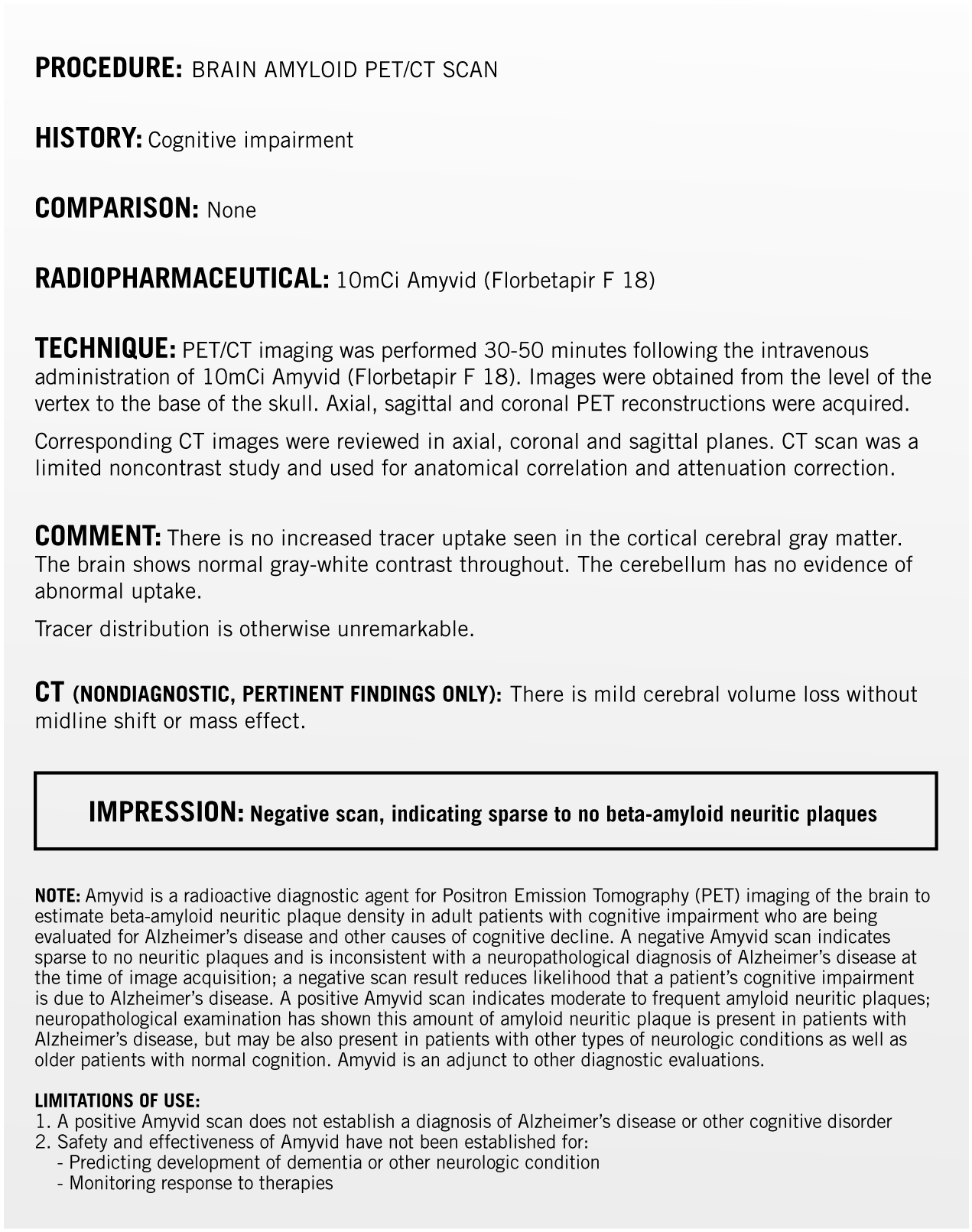
Amyvid is indicated for Positron Emission Tomography (PET) imaging of the brain to estimate beta-amyloid neuritic plaque density in adult patients with cognitive impairment who are being evaluated for Alzheimer's Disease (AD) and other causes of cognitive decline. A negative Amyvid scan indicates sparse to no neuritic plaques and is inconsistent with a neuropathological diagnosis of AD at the time of image acquisition; a negative scan result reduces the likelihood that a patient's cognitive impairment is due to AD. A positive Amyvid scan indicates moderate to frequent amyloid neuritic plaques; neuropathological examination has shown this amount of amyloid neuritic plaque is present in patients with AD, but may also be present in patients with other types of neurologic conditions as well as older people with normal cognition. Amyvid is an adjunct to other diagnostic evaluations. Limitations of Use:
Amyvid for intravenous use is supplied in multidose vials containing |